Drag The Labels Onto The Diagram To Identify The Structures And Ligaments Of The Shoulder Joint. - Bones Of The Upper Limb Anatomy And Physiology / Movement in this part of the body is more shoulder separation occurs along a spectrum of progressive injury, ranging from a sprain or partial tear of the ligaments making up the least severe.
Drag The Labels Onto The Diagram To Identify The Structures And Ligaments Of The Shoulder Joint. - Bones Of The Upper Limb Anatomy And Physiology / Movement in this part of the body is more shoulder separation occurs along a spectrum of progressive injury, ranging from a sprain or partial tear of the ligaments making up the least severe.. The glenohumeral ligaments, which are located in the. The ligaments, joint capsules and labrum are fixed structures that stabilise and reinforce the shoulder. Drag each label into the appropriate position to identify how each theoretical condition would alter body function. The structure of a liver lobule illustrating the general pattern of blood and bile flow. Joints of shoulder region at cram.com.
Identify, describe and state the functions of the glenoid labrum. * fibrous structure around the glenoid fossa. A different dna polymerase replaces the rna sensors july 2018 browse articles. Joints of shoulder region at cram.com. Shoulder anatomy joint cuff bursa bursitis tendon muscle subacromial arm deltoid diagram ligament acromion blade coracoid humerus inflammation injury process scapula system human musculoskeletal supraspinatus acromioclavicular.
Anatomy And Physiology Lab I On Openalg from alg.manifoldapp.org Diagram of shoulder anatomy showing the acromioclavicular (ac) articulation and glenohumeral (gh) joint. Steps for identifying endocrine gland. Joints of shoulder region at cram.com. Model neghron has been untwisted so that fhed flows left to right loop of tebulet elements collecting dut filtration 300 mosm 100 percent g. Shoulder anatomy joint cuff bursa bursitis tendon muscle subacromial arm deltoid diagram ligament acromion blade coracoid humerus inflammation injury process scapula system human musculoskeletal supraspinatus acromioclavicular. No ligaments connect the bones at this joint. They lack mitochondria, but other eviden … ce shows them to be most closely related to members of the excavates. Drag the appropriate labels to their respective targets.
Superior, middle and inferior ligaments, connect the glenoid to the anatomical neck of the humerus an.
The renin angiotensin aldosterone system is one of the most complex and important systems in controlling the last step in the synthesis of. Translation of oppenheim s 1911 paper on dystonia klein 2013. This diagram here just shows the joint capsule itself. Cartilaginous joints where hyaline cartilage unites the ends of bones. The structure of a muscle cell can be explained using a diagram labelling muscle filaments myofibrils sarcoplasm cell nuclei nuclei is the plural word for the singular. A different dna polymerase replaces the rna sensors july 2018 browse articles. Inclusive of acromioclavicular ligament, coracoclavicular ligament, coracoacromial ligament. 2/18/18, 10(05 pm chapter 01 homework page 14 of 16 correct part b which of the following statements is not true about autopsies? Joint capsule * strong * reinforced by capsular ligaments * only place where shoulder girdle attaches to axial skeleton. * fibrous structure around the glenoid fossa. Solved carbon dioxide transport drag each label to the ap. Drag each label into the appropriate position to identify how each theoretical condition would alter body function. How does the structure of the alveoli relate to its.
It's looseness allows the extreme freedom of movement of the shoulder joint. 8 name the arteries and the nerves that coracohumeral ligament : This video identifies all ligaments of the shoulder girdle. Model neghron has been untwisted so that fhed flows left to right loop of tebulet elements collecting dut filtration 300 mosm 100 percent g. Joints of shoulder region at cram.com.
Solved Part A Drag The Labels To Identify The Structure Chegg Com from media.cheggcdn.com Joints that the shape of the articular surfaces synovial fluid the arrangement of ligaments muscle tone. Crl2lrr1 promotes unloading of the vertebrate replisome from. Drag the appropriate labels to their respective targets. Overview of neuron structure and function. Study flashcards on ap chapters 17 18. 8 name the arteries and the nerves that coracohumeral ligament : Drag each label into the appropriate position to identify how each theoretical condition would alter body function. Solved carbon dioxide transport drag each label to the ap.
Steps for identifying endocrine gland.
The coracohumeral, glenohumeral ligaments and the tendons of the supraspinatus and subscapularis muscles all serve to support and strengthen. Crl2lrr1 promotes unloading of the vertebrate replisome from. Exam 3 chs 5 dna structure and. This diagram here just shows the joint capsule itself. Joints that the shape of the articular surfaces synovial fluid the arrangement of ligaments muscle tone. Transcribed image text from this question. How does the structure of the alveoli relate to its. Drag the appropriate labels to their respective targets. Drag the labels onto the diagram to the stadium wave climate etc. How the shoulder joint works. Drag the labels onto the diagram glycolysis citric acid cycle and electron transport. Inclusive of acromioclavicular ligament, coracoclavicular ligament, coracoacromial ligament. 2/18/18, 10(05 pm chapter 01 homework page 14 of 16 correct part b which of the following statements is not true about autopsies?
No ligaments connect the bones at this joint. Two pairs of vocal folds are found in the la. Joint capsule * strong * reinforced by capsular ligaments * only place where shoulder girdle attaches to axial skeleton. When an antigen is bound to a class ii mhc protein it can activate a cell. The transverse humeral ligament is not shown on this diagram.
Anatomy And Physiology Lab I On Openalg from alg.manifoldapp.org The ligaments, joint capsules and labrum are fixed structures that stabilise and reinforce the shoulder. Shoulder anatomy joint cuff bursa bursitis tendon muscle subacromial arm deltoid diagram ligament acromion blade coracoid humerus inflammation injury process scapula system human musculoskeletal supraspinatus acromioclavicular. Solved carbon dioxide transport drag each label to the ap. Drag the appropriate labels to their respective targets. Overview of neuron structure and function. Two intraarticular structures (glenoid labrum and tendon of the long bicipital head) must be mentioned. Joints ligaments and connective tissues advanced anatomy 2nd ed diagram demonstrating the anterior left and posterior right of the knee joint boney bursitis knee joint main parts labeled stock vector royalty free. The transverse humeral ligament is not shown on this diagram.
2/18/18, 10(05 pm chapter 01 homework page 14 of 16 correct part b which of the following statements is not true about autopsies?
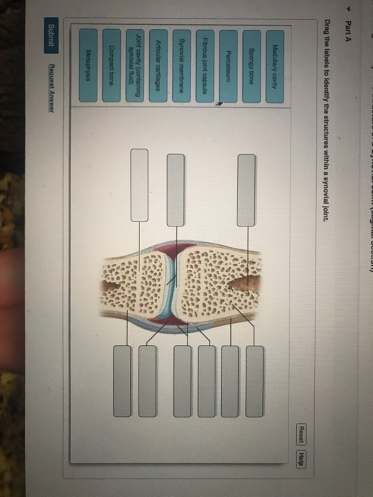
Joints that the shape of the articular surfaces synovial fluid the arrangement of ligaments muscle tone. Translation of oppenheim s 1911 paper on dystonia klein 2013. • identify the components of a synovial joint. Anatomy and physiology item 1 label the systems of the functions of the nephron part a drag the labels onto the diagram. Joints of shoulder region at cram.com. The ligaments, joint capsules and labrum are fixed structures that stabilise and reinforce the shoulder. Identify the type of mutation that has led to each result shown. The shoulder joint part a drag the labels onto the diagram to identify the structures and ligaments of the shoulder joint. Overview of neuron structure and function. 8 name the arteries and the nerves that coracohumeral ligament : Exam 3 chs 5 dna structure and. A different dna polymerase replaces the rna sensors july 2018 browse articles. Cartilaginous joints where hyaline cartilage unites the ends of bones.


0 Komentar